Introduction: The lateral skull base is one of the most anatomically complex regions of the human body, owing to the proximity of vital neurovascular structures. Knowledge of anatomical relations is essential for the skull base surgeon in operative planning and in preventing inadvertent damage and surgical morbidity. Furthermore, fascial planes in this region can provide safe routes of access and act as barriers to spread of malignancy. Although the fascial layers and spaces of the head and neck have been well described in literature, a study from a lateral approach has not been conducted.
Aim: To examine the critical anatomical relationships and fascial layers relevant to lateral approaches to the skull base.
Methods: Five soft-embalmed cadaveric heads (ten sides) were dissected from a lateral approach to reveal the layers of the deep cervical fascia, together with their attachments and relations.
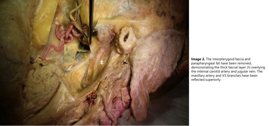
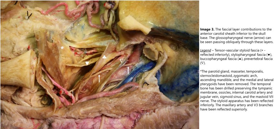
Results: All three layers of the deep cervical fascia contributed to the fascial network of the lateral skull base. Anteriorly, the most cranial part of the carotid sheath was formed by multiple fused fascial layers, with contributions from the tensor-vascular styloid, stylopharyngeal, buccopharyngeal, and prevertebral fascias. This fused thick layer protected the underlying carotid artery, jugular vein, and lower cranial nerves. The glossopharyngeal nerve pierced this sheath on average 48mm below the skull base.
Conclusions: A complex network of fascial planes exists in the lateral skull base region, whereby multiple layers fuse to form protective coverings for neurovascular structures. Understanding these layers and anatomical relations can help the surgeon plan and safely perform challenging procedures in this complex region.
.jpg)